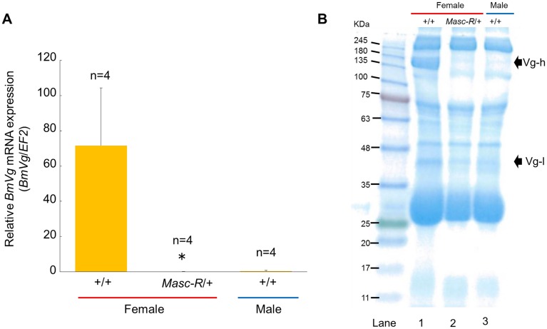
Fig 4. Expression of Masc-R severely diminished vitellogenin synthesis in the female fat body.
(A) Quantification of the BmVg mRNA level within 3 hours after pupation using qRT-PCR. EF2 served as an internal standard. Error bar: SD; * significant differences at the 0.05 level (Welch’s t-test) compared with the +/+ female. (B) SDS-PAGE analysis of the whole hemolymph within 3 hours after pupation. Hemolymph sample applied in each lane was a mixture of hemolymph collected from three individuals. The arrows indicate the protein bands corresponding to the molecular weight of BmVg heavy chain (Vg-h) and BmVg light chain (Vg-l), respectively.

