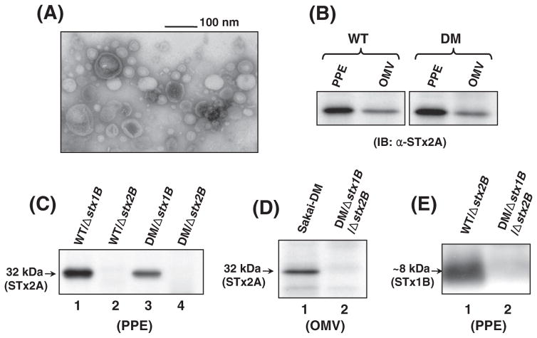
Fig. 1.
TEM and IB analyses for characterization of the ΔstxB mutants. (A) TEM visualization of OMVs from Sakai-DM/Δstx2B mutant. The image showed typical sizes of OMVs ranges 20–100 nm in diameter. (B, C, and D) Immunoblots for detection of STx2A subunit in the PPE and OMV samples. STx2A was identified in the periplasm and OMVs of the parental O157 strains (B) by using anti-STx2A mAb, however, it was absent in the PPE samples (lanes 2 and 4, C) and the OMVs (lane 2, D) of the Δstx2B mutants. (E) STx1B subunit was probed with anti-STx1B mAb in the periplasm of the Δstx2B mutant (lane 1) but was absent in the Δstx1B mutant (lane 2).

