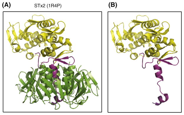
Fig. 5.
Ribbon structure of STx2 toxin. Structures of STx2 holotoxin (A) and STx2A (B) were obtained from the Protein Data Bank (accession code: 1R4P) and displayed after manipulation by using the PyMOL Molecular Graphics System, Version 1.3, Schrödinger, LLC. The 32 kDa STx2A subunit, subdivided as A1 (yellow) and A2 (magenta) fragments in the panel A, is known to be non-covalently associated with five STx2B (7.7-kDa) subunits (green). In the absence of the B-pentamer (panel B), the A2-domain (magenta) of STx2A would be then exposed to periplasmic proteases. (For interpretation of the references to color in this figure legend, the reader is referred to the web version of this article.)

