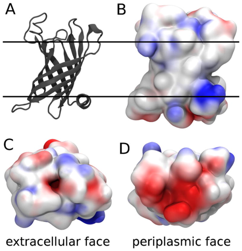
Fig. 1.

The Gram negative bacterial outer membrane enzyme PagP. (A) Cartoon representation of the crystal structure of PagP in LDAO (PDB ID 1THQ) (Ahn et al., 2004). The proposed (Ahn et al., 2004) bilayer interfaces are depicted as solid lines. (B–D) Electrostatic potential of PagP, computed by the adaptive Poisson-Boltzmann solver (Baker et al., 2001), mapped onto its van der Waals surface by VMD (Humphrey et al., 1996) showing (B) side view along the proposed (Ahn et al., 2004) bilayer plane, and (C) extracellular and (D) periplasmic apical surfaces. (Red) Electronegative and (blue) electropositive regions of the protein surface are highlighted. For interpretation of the references to color in this figure legend, the reader is referred to the web version of the article.
