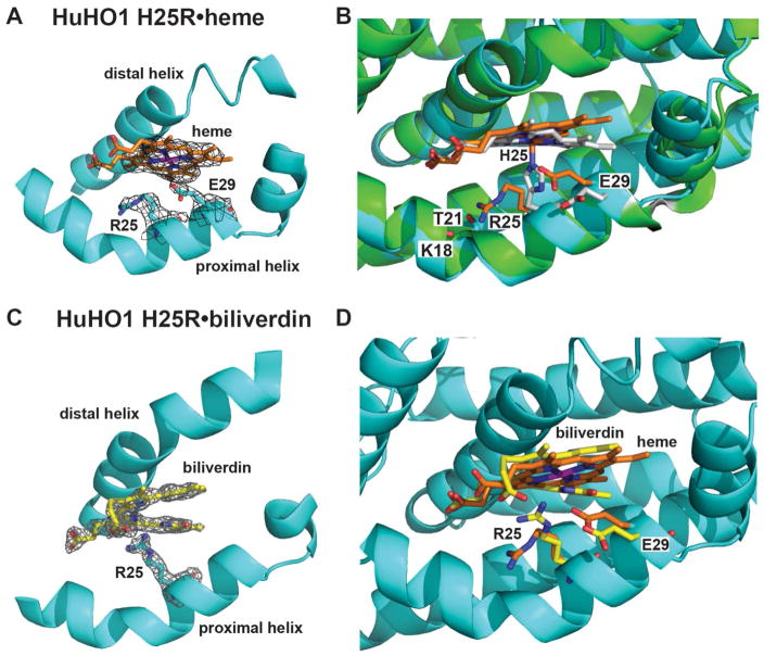
Figure 7. X-ray structural studies of HuHO1 H25R.
(A) Sigma-A-weighted 2Fo − Fc electron density map (contoured at 1σ) from the 2.95-Å resolution structure of heme (orange) bound to HuHO1 H25R (determined herein, PDB 4WD4). (B) Superposition of the HuHO1 H25R•heme structure (protein in cyan, heme in orange) and the previously published WT HuHO1•heme structure (PDB 1N45, protein backbone in green, side-chains in white, heme in white). (C) Sigma-A-weighted 2Fo − Fc electron density map (contoured at 1.2σ) from the 2.08-Å resolution structure of biliverdin IXα (yellow) bound to HuHO1 H25R (determined herein, PDB 5BTQ). (D) Superposition of the heme- and biliverdin-bound structures of HuHO1 H25R (heme in orange, biliverdin in yellow, residues from heme-bound structure in orange, residues from BV-bound structure in yellow). See Table S1 for data acquisition and refinement statistics

