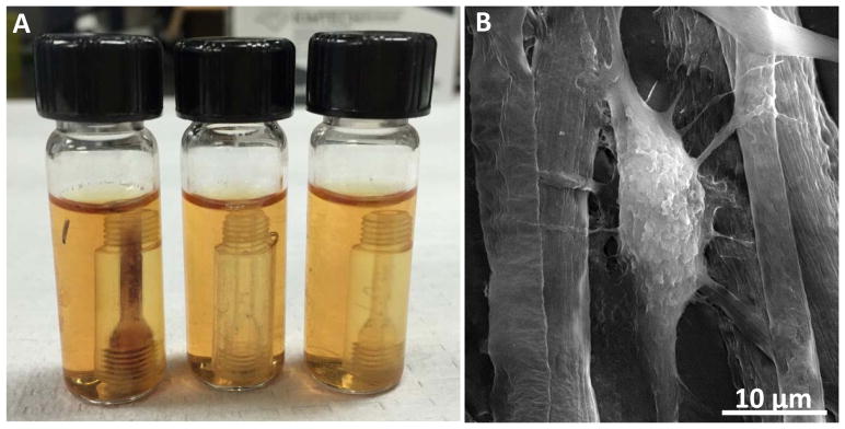
Figure 4.
(A) MTS assay to quantify viable HDFs on different devices. The three devices from left to right are: a PCL fiber coated device, a bare device (no fibers coated), and a PCL layer coated device. The same HDF seeding and culture process was conducted on all three devices. A layer of dark purple was observed on the inside of the channel of the left device, which indicated viable cells were adhering on the fibers coated on the channel. The solution from the vials was pipetted into a 96-well plate for absorption measurements, which revealed that 4.13 × 10⁵ (± 0.76 × 10⁵) cells were cultured on such a device. The middle and the right devices, however, did not show color change in the channels, indicating no cells were adhered onto the inner walls of the devices. (B) SEM image of an HDF cultured on the coated fibers in a fluidic device. Some pseudopodia were formed by the cell to attach surrounding fibers, which further confirmed that HDFs can adhere on the fibers

