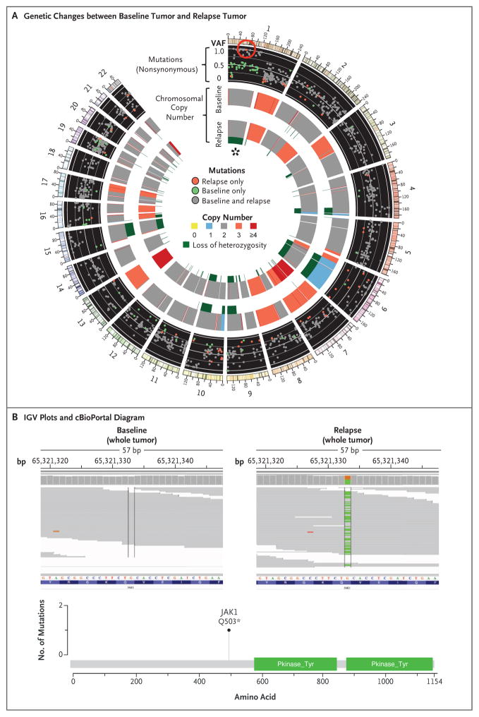
Figure 2. Acquired JAK1 Loss-of-Function Mutation at Relapse, with Accompanying Loss of Heterozygosity.
In Panel A, a Circos plot25 of Patient 1 shows differences in whole-exome sequencing between the pre-pembrolizumab and post-relapse biopsies. The red circle highlights a new, high-allele-frequency, relapse-specific mutation in the gene encoding Janus kinase 1 (JAK1) in the context of chromosomal loss of heterozygosity (asterisk). Each wedge represents a chromosome. In the outer track (black background), each point represents a nonsynonymous mutation, with most detected in both biopsy samples (gray) rather than at relapse only (red) or baseline only (green). The y-axis position indicates the variant allele frequency (VAF) at relapse, unless baseline-specific. The middle and inner tracks show copy-number status for the baseline and relapse biopsy, respectively; dark green in the subtrack indicates loss of heterozygosity. In Panel B, Integrative Genomics Viewer (IGV) plots (top) show that the JAK1 Q503* nonsense mutation is relapse-specific, and the cBioPortal26 diagram (bottom) shows that the JAK1 mutation is upstream of the kinase domains.

