
Figure 2. NCI/ADR-RES spheroid growth.

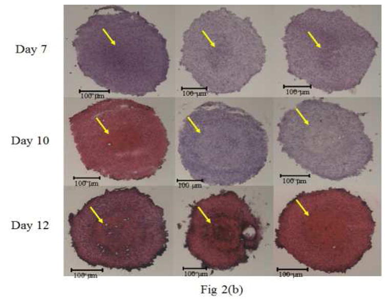
(a) Spheroid diameter with age (Mean ± S.D., n=3) (b) H&E stained sections of NCI/ADR-RES spheroids by fluorescence microscopy. From day 7 onward, the formation of a distinctly stained core (indicated by arrow) became visible. Day 10 spheroids showed 2 distinct layers consisting of a proliferative outer shell and the necrotic inner core. By day 12, the spheroids had begun shedding cells on the periphery.