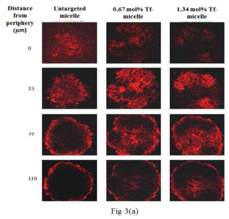
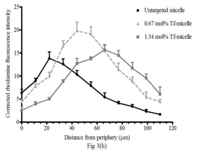
Figure 3. Micelle penetration of spheroids.

(a) Z-stack images taken by confocal microscopy showing penetration of untargeted and Tf-targeted rhodamine (Rh)-labeled micelles in 4 days old NCI/ADR-RES spheroids. The images are of spheroid sections at 0, 33, 77 and 110 μm distance from the periphery. The red color indicates the presence of Rh-labeled micelles. The micelle association with the deeper layers of the spheroid was higher when targeted. (b) The corrected rhodamine fluorescence intensity represents the micelle association of targeted and untargeted micelles in NCI/ADR-RES spheroids from the periphery to the inner layers.

