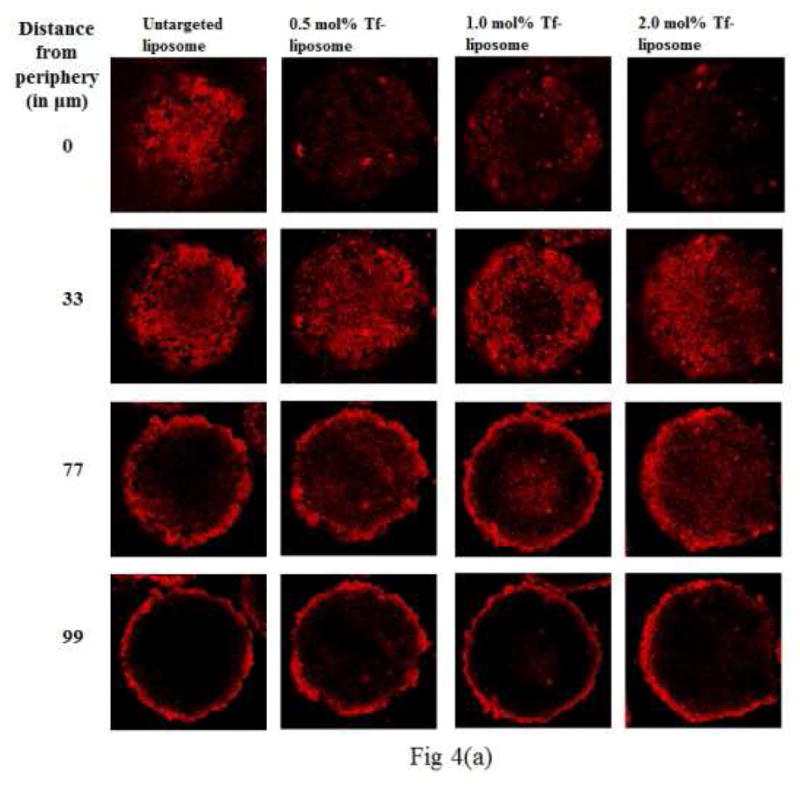
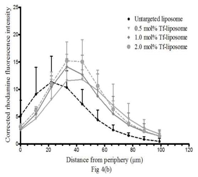
Figure 4. Liposome penetration in spheroids.

(a) Z-stack images taken by confocal microscopy show penetration of untargeted and Tf-targeted rhodamine (Rh)-labeled liposomes in 4 days old NCI/ADR-RES spheroids. The images are of spheroid sections at 0, 33, 77 and 99 μm distance from the periphery. The red color indicates the presence of Rh-labeled liposomes. The association with the deeper layers of the spheroid was higher with Tf-targeted liposomes. (b) The corrected rhodamine fluorescence intensity vs distance from the periphery quantitates the liposome association of targeted and untargeted liposomes in NCI/ADR-RES spheroids.

