Figure 3.
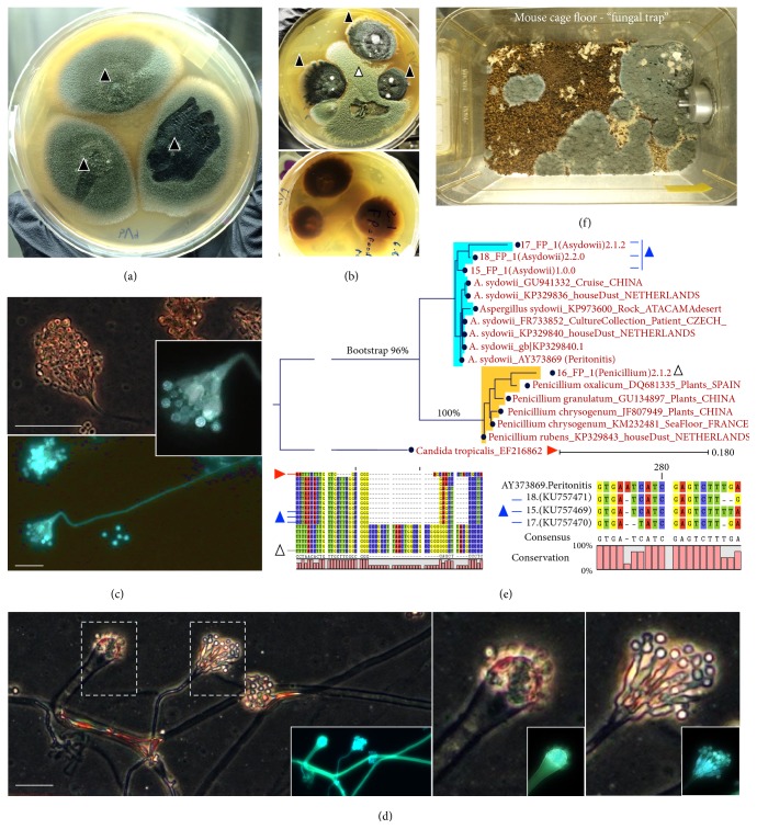
Culture and molecular identification of Aspergillus sydowii. (a) A. sydowii in potato dextrose agar supplemented with gentamicin and chloramphenicol (30 and 125 mg/L) with typical blue-green colonies (solid triangles, three distinct isolates from GF-irradiated rodent feed in two GF isolators). (b) Coculture with a Penicillium feed isolate (open triangle) to emphasize colony morphology and pigment production by A. sydowii (transagar pigmentation). (c) Adhesive tape preparation of A. sydowii colony to illustrate conidiophores, conidia, and typical fruiting head vesicle arrangement with nonseptate conidiophore (400x; phase-contrast microscopy on Calcofluor White from Remel; Alexa Fluor 430; emission 541; absorption 433 nm; scale bar, 30 μm). (d) See distinct types of conidiophore formation by A. sydowii. (e) Neighbor-Joining phylogram of A. sydowii isolates in the context of other published 18S internal transcribed spacers ITS1–4 sequences and Penicillium spp. Notice the close-up of region where strains have sequence dissimilarity. (f) Panoramic photo of mouse cage bedding (set as “fungal trap”; i.e., mouse cage incubated for 3 weeks at 23°C) at the end of clinical observation period confirming mice were still exposed or shedding A. sydowii.

