
Figure 1.
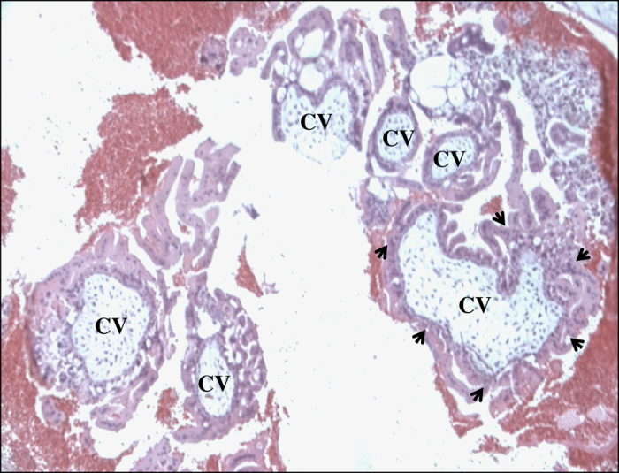
Photomicrograph of a complete hydatidiform mole (CHM) from patient 1448 showing several chorionic villi (CV). Circumferential trophoblastic proliferation around one villous on the right bottom is indicated by arrows.
Official websites use .gov
A
.gov website belongs to an official
government organization in the United States.
Secure .gov websites use HTTPS
A lock (
) or https:// means you've safely
connected to the .gov website. Share sensitive
information only on official, secure websites.
Photomicrograph of a complete hydatidiform mole (CHM) from patient 1448 showing several chorionic villi (CV). Circumferential trophoblastic proliferation around one villous on the right bottom is indicated by arrows.