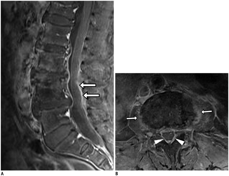
Fig. 11. Pyogenic (staphylococcal infection) spondylodiscitis.
73-year-old woman with mild fever and back pain. Fat-suppressed, contrast-enhanced sagittal T1-weighted imaging (T1WI) showed only thin linear enhancement of epidural space (arrows) without signal changes in bone marrow or cortical changes (A). However, fat-suppressed, contrast-enhanced axial T1WI (B) showed faint paraspinal (arrows) and epidural (arrowheads) enhancement. Two months later, follow-up MRI (not shown) demonstrated aggravation of infectious process.

