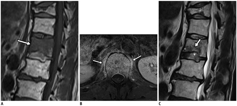
Fig. 13. Acute compression fracture with bone marrow edema mimicking infection.
62-year-old man with history of medication for osteoporosis. Sagittal T1-weighted imaging (T1WI) (A) revealed L1 vertebral body with subchondral bone defect at its lower endplate with marked bone marrow edema (arrow). Fat-suppressed, contrast-enhanced axial T1WI (B) depicted paraspinal soft tissue edema (arrows). Sagittal T2-weighted imaging (C) exhibited faint fracture line (arrow) with low signal intensity, highly suspicious for compression fracture.

