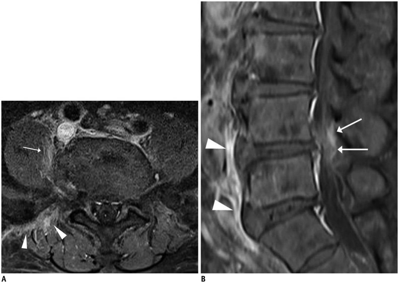
Fig. 16. Pyogenic spondylodiscitis caused by procedure.
68-year-old male patient presented with lower back pain and fever 3 days after selective nerve root block. Fat-suppressed, contrast-enhanced axial T1-weighted imaging (T1WI) (A) showed mild inflammatory changes in right paraspinal soft tissue (arrow) and back muscle area (arrowheads) corresponding to needle track. Fat-suppressed, contrast-enhanced sagittal T1WI (B) showed faint epidural enhancement (arrows) and linear subligamentous enhancement (arrowheads). MRI (not shown) obtained 10 days later showed aggravation of infectious process.

