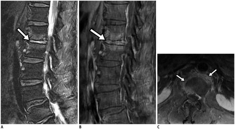
Fig. 3. Pyogenic spondylodiscitis.
68-year-old man with lower back pain. Fat-suppressed sagittal T2-weighted image (A) showed bright high signal intensity (arrow) of L1–2 disc, which was well enhanced (arrow) on fat-suppressed, contrast-enhanced sagittal T1-weighted imaging (B). Fat-suppressed, contrast-enhanced axial T1-weighted image (C) demonstrated enhancement of paraspinal soft tissue and peripheral portion of disc (arrows).

