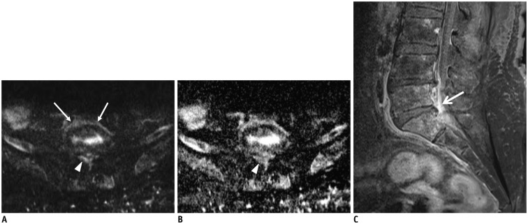
Fig. 6. Pyogenic spondylodiscitis with epidural abscess.
72-year-old woman with lower back pain and underlying hepatocellular carcinoma. Axial diffusion-weighted imaging (A) showed high signal intensity in paraspinal area (arrows) and epidural area (arrowhead) at L5–S1 level. Epidural lesion demonstrated signal loss (arrowhead) on axial apparent diffusion coefficient (ADC) map (B) and had lower ADC value (0.93 x 10⁻³ mm2/s). Fat-suppressed, contrast-enhanced sagittal T1-weighted imaging (C) showed marked epidural enhancement (arrow) at L5–S1 level.

