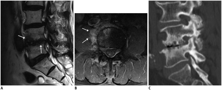
Fig. 9. Type 1 Modic change.
66-year-old man with history of lung cancer and lower back pain. Findings of multifocal cortical discontinuities or erosions of endplates (arrows) on sagittal T2-weighted imaging (A) and paraspinal soft tissue enhancement (arrows) on fat-suppressed, contrast-enhanced axial T1-weighted imaging (B) were confused with infection. However, reformatted sagittal CT (C) findings of vacuum disc, well-defined sclerosis, and erosions of vertebral endplates reduced possibility of infection.

