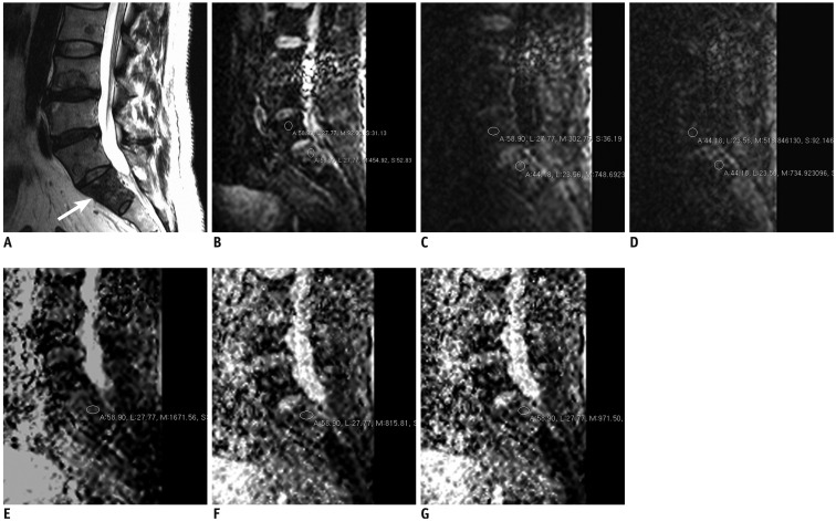
Fig. 2. 58 years old man with oropharyngeal carcinoma metastasis.
A. 3T MRI T2-weighted sagittal image (TR/TE, 2500/120) shows metastatic lesion in S1 (arrow). B. Diffusion weighted image with b value of 0. Regions of interest were placed within lesion (lower circle) and normal marrow (upper circle) of vertebral body. C. Diffusion weighted image with b value of 400. D. Diffusion weighted image with b value of 1000. E. ADC map with b value of 0–400. Region of interest were placed within lesion. F. ADC map with b value of 0–1000. G. ADC map with b value of 0–400–1000. First, both readers diagnosed lesion as malignant based on routine MR images. During second session, both readers diagnosed lesion as malignant based on routine MR images with additional information of DWI with b value 400. During third session, readers (both reader 1 and reader 2) diagnosed lesion as malignant based on routine MR images with additional information of DWI with b value 1000. ADC = apparent diffusion coefficient, DWI = diffusion weighted image, TE = echo time, TR = repetition time

