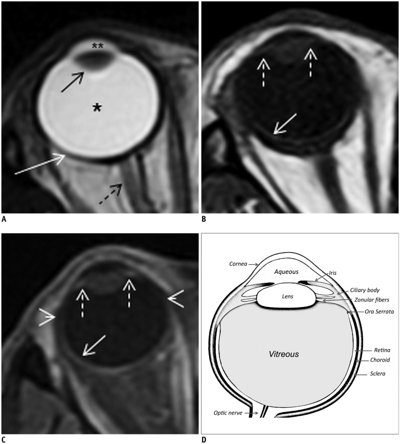
Fig. 1. Normal globe anatomy on orbital MRI.
Lens (black arrow) and sclera (white arrow) show hypointense signal on all sequences. A. On axial T2W images, vitreous (*) and aqueous humour in anterior chamber (**) are diffusely hyperintense. Optic nerve is labeled (dashed black arrow). B. Axial T1W image of right globe. Retina and choroid appear as single hyperintense layer (white arrow) with enhancement on fat-saturated post contrast T1W image (C, white arrow). Ciliary bodies form part of choroid (dashed white arrows, B, C). Approximate position of ora serrata is shown (small white arrowheads). D. Anotated illustration of globe for comparison with MRI anatomy. T1W = T1-weighted, T2W = T2-weighted

