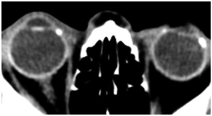
Fig. 10. Axial non-contrast image from brain CT assessment of altered mental state shows bilateral lens prostheses with incidental scleral calcifications at insertion of medial rectus on right and both medial and lateral recti on left.
These calcifications represent normal part of aging. Scleral bands would appear more linear, as compared to punctate calcifications observed.

