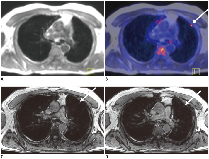
Fig. 4. 62-year-old male patient with thyroid cancer.
A. Dixon sequence in expiration. B. Fused PET/Dixon. C. VIBEin. D. VIBEex. In fused images, focal uptake was observed in periphery of left lung (arrow), which was rated as lesion based on PET information. In Dixon sequence, there was no correlate to finding while in VIBEin and VIBEex small subpleural nodule was visible, which was rated as potentially malignant with confidence level of 1 (= highly likely). In follow-up imaging, lesion grew in size and was rated as metastasis in standard of reference. PET = positron emission tomography, VIBEex = volume interpolated breath-hold examination acquired in expiration, VIBEin = volume interpolated breath-hold examination acquired in inspiration

