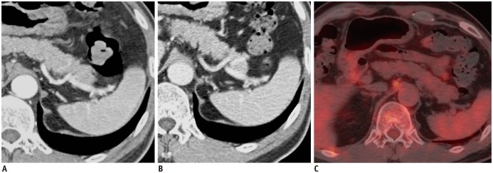
Fig. 2. 62-year-old man with diffuse large B-cell lymphoma.
Axial contrast-enhanced MDCT shows obliteration of normal heterogeneous enhancement of spleen (ONHES) on AP image (A) and homogeneous enhancement on PVP image (B) with 247 cm3 of mean splenic index. However, there is no evidence of increased FDG uptake in spleen, suggesting false-positive finding for ONHES (C). AP = arterial phase, FDG = fluorodeoxyglucose, MDCT = multidetector CT, PVP = portal venous phase

