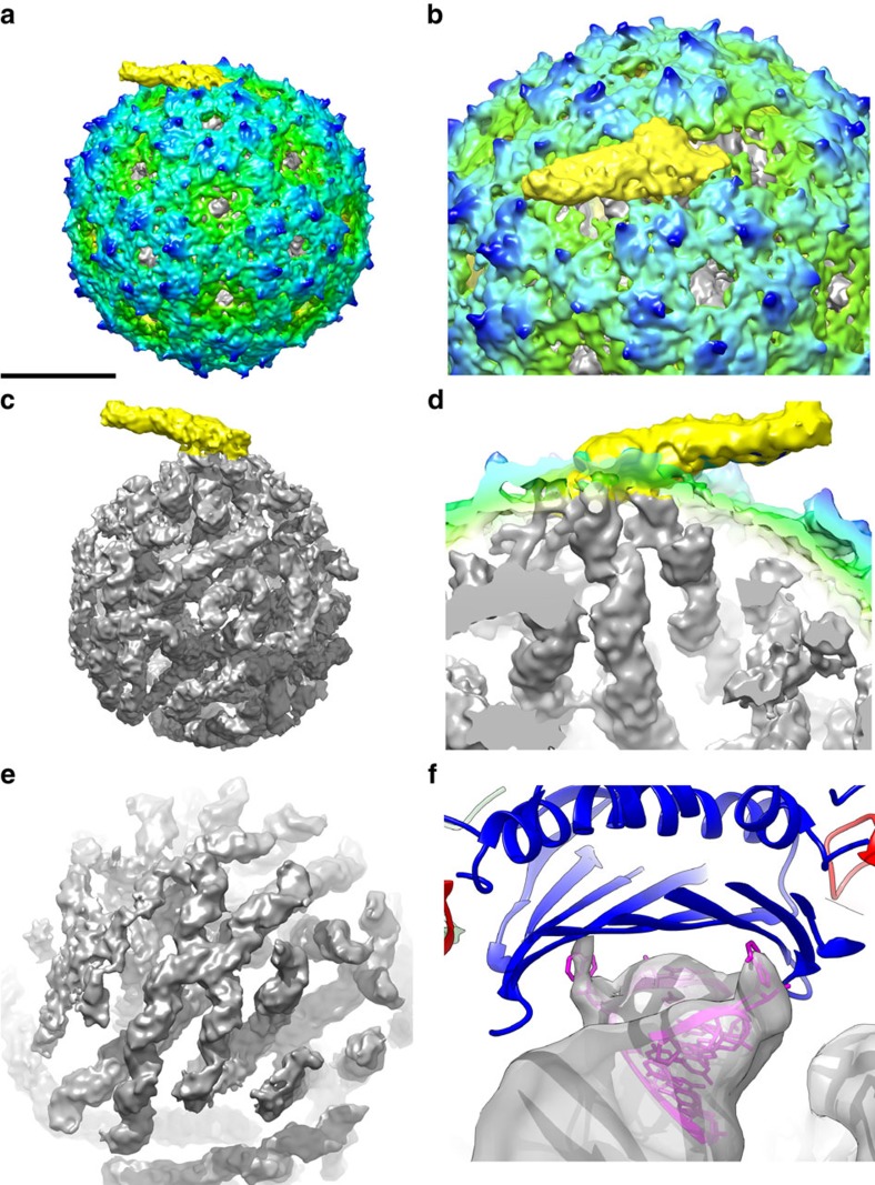
Figure 1. The asymmetric reconstruction of bacteriophage MS2.
Asymmetric structure of bacteriophage MS2 (green–blue radially coloured) shows the AP (yellow) (a), which replaces one CP dimer (b). Inside the protein capsid a structured genome (grey) is present (c) that is connected to the AP (d). The reconstruction shows the double-stranded helices in the stem loop structures (e). At some positions individual NA's connecting to the capsid are resolved, as shown by fitting of the X-ray structure of the 19-nucleotide TR (magenta) bound to the capsid (blue) (pdb:1ZDH) in the EM density (grey) (f). Scale bar is 100Å.

