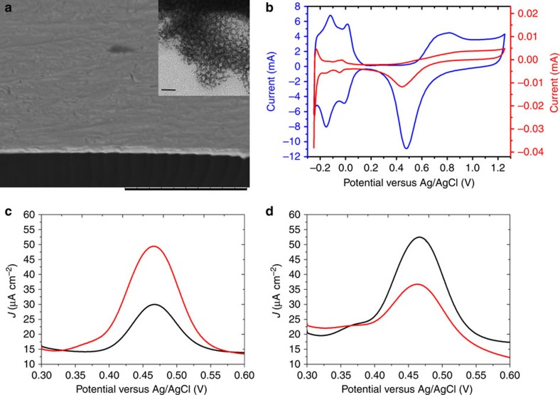
Figure 2. Characterization of chiral-imprinted mesoporous platinum.
(a) Scanning electron microscopy image of a typical cross-section of the metal film. Scale bar, 10 μm. Inset:TEM image of the mesopores with a scale bar of 20 nm. (b) Cyclic voltammograms of flat-polished platinum (red) and a chiral-imprinted mesoporous platinum film (blue), recorded in 0.5 M H2SO4 at 100 mV s−1. (c,d) Differential pulse voltammograms in 4 mM D-DOPA (black) and L-DOPA (red) (50 mM HCl as supporting electrolyte) of chiral mesoporous platinum electrodes imprinted with (R)- and (S)-MA, respectively.

