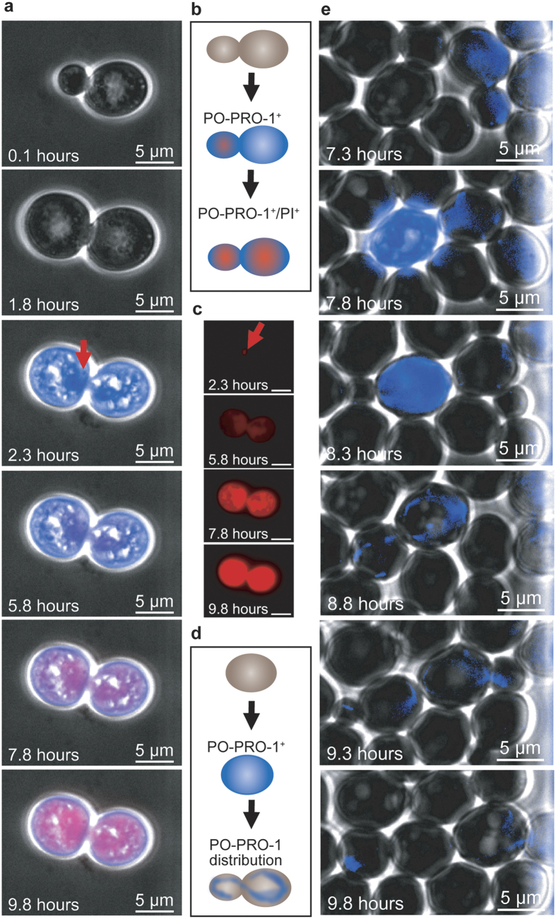
Figure 5. Budding and cell death.
(a) A budding yeast cell was injured at the budding neck (marked with red arrow) and subsequently died. (b) Schematic drawing of an apoptotic mother cell that lost its membrane potential at the same time as its bud cell, which was injured near the budding neck. Both cells were PO-PRO-1+/PI+. (c) The pictures indicate that spatiotemporally resolved PI fluorescence diverges from the merged images in (a). (d) Schematic drawing of cell recovery due to cell budding. The cell became PO-PRO-1+ after membrane potential loss. The cell eventually initiated PCD while undergoing replication and proceeded with budding and actin-assisted DNA distribution (AT-rich regions appear fluorescent blue). (e) A cell exhibiting apoptotic-like behaviour followed by recovery due to budding is marked with white arrows. PO-PRO-1 fluorescence increased and was maintained for one hour before budding was initiated. DNA passage into the daughter cell was observable due to the presence of the DNA indicator PO-PRO-1. Mother and daughter yeast cells proceeded to budding followed by the dilution of fluorescence in the next filial generation.

