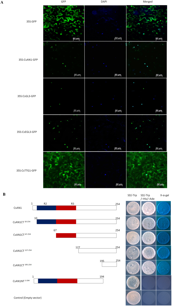
Figure 4. Subcellular localizations of CsAN1, CsGL3, CsEGL3 and CsTTG1 and transactivation assay of CsAN1.
(A) Subcellular localization of CsAN1, CsGL3, CsEGL3 and CsTTG1 in epidermal cells of N. benthamiana leaves. GFP fluorescence was detected 50 hours after infiltration. The nuclei were indicated by DAPI staining. (B) Transactivation assay of CsAN1. Either the full length or a truncated ORF of CsAN1 was fused with pGBKT7, and transformed yeasts were selected on SD/-Trp or SD/-Trp/-His/-Ade/X-α-gal media for 3 d at 30 °C. Transcription activation was monitored by the detection of yeast growth and an α-galactosidase assay.

