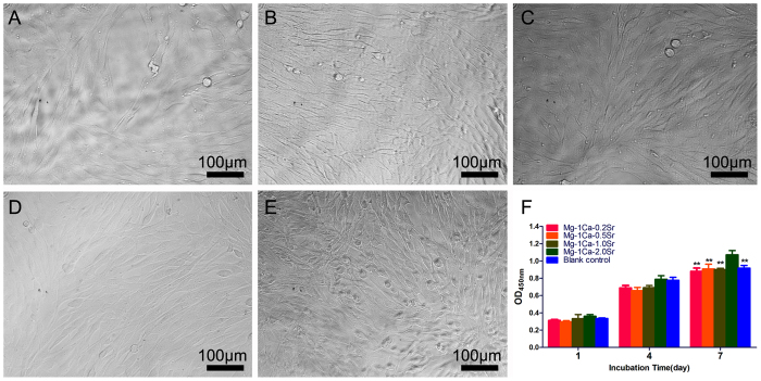
Figure 5. Cell viability assay in four alloys extracts after 1, 4 and 7 days incubation was measured by colorimetric CCK-8 assay.
Cells cultured in α-MEM medium containing 10% FBS were set as control group. Cell morphology was observed by inverted phase contrast microscope on day 4, and the images showed that cells in different extracts were normal and healthy (A) Mg-1Ca-0.2Sr alloy; (B) Mg-1Ca-0.5Sr alloy; (C) Mg-1Ca-1.0Sr alloy; (D) Mg-1Ca-2.0Sr alloy), similar to that of the blank control (E). CCK-8 assay (F) showed that the absorbance of cells cultured in four Mg alloys extracts medium was gradually increased. At day 7, cell number of Mg-1Ca-2.0Sr alloy group was slightly higher than other group and reached the peak (p < 0.01). All data represent the mean ± standard deviation of three independent experiments. **p < 0.01 compared with Mg-1Ca-2.0Sr alloy group.

