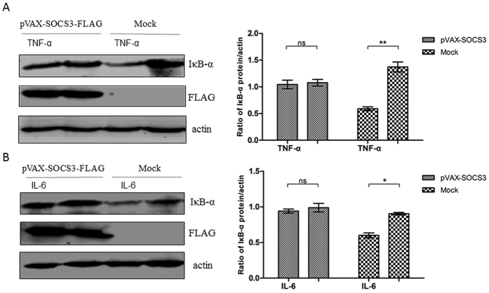
Figure 6. SOCS3 inhibits the induction of NF-κB signaling by TNF-α and IL-6 in vitro.
PK-15 cells were transfected with pVAX-SOCS3 (1 μg) or pVAX (1 μg). At 24 h post-transfection, the cells were treated with TNF-α (50 ng/ml) or IL-6 (200 ng/ml) for 20 min; non-stimulated cells served as a control. Then, the cells were lysed and subjected to western blotting to analyze IκB-α degradation following stimulation with (A) TNF-α or (B) IL-6. IκB-α protein expression is presented relative to that of β-actin. Statistical data were analyzed by one-way analysis of variance (*P < 0.05; **P < 0.01; and ***P < 0.001; ns, not significant (P > 0.05)). All data are expressed as the mean ± SD. The samples were derived from the same experiment, and the blots of IκB-α and actin were processed in parallel in the same gel. The blots of SOCS3-FLAG were performed in another gel using the same samples because the bands of SOCS3-FLAG and IκB-α were too close to differentiate. The samples for blotting were prepared at the same concentrations, and the two gels were run under the same conditions.

