-
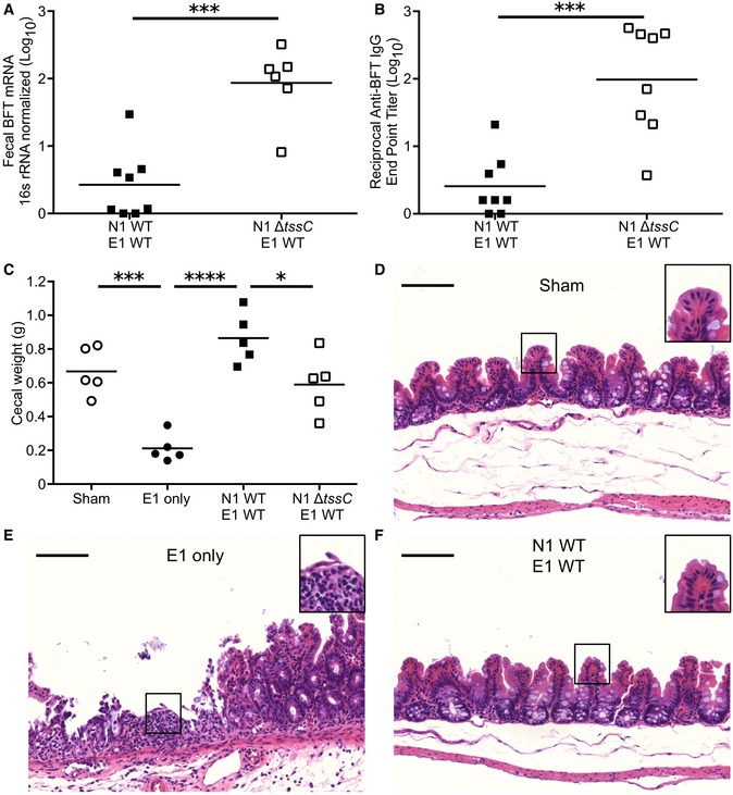
A
Mice were co‐colonized with E1 and either N1 WT (n = 4) or N1 ΔtssC (n = 3). Five days post‐inoculation, fecal RNA was extracted and tested for BFT expression via qRT–PCR.
-
B
Four weeks after co‐colonization with E1 and either N1 WT or N1 ΔtssC (n = 4 mice per group), the sera were collected, tested via ELISA for anti‐BFT IgG, and endpoint titer calculated.
-
C–F
Mice pre‐treated with DSS were inoculated with no organisms (sham), E1 only, or E1 competed with N1 WT or N1 ΔtssC. Five days post‐inoculation, the ceca were weighed (C) and fixed for histopathological examination after sham (D), E1 only (E) and E1‐N1 WT (F) colonizations. Scale bars denote 100 μm (main image) and 200 μm (inset).
Data information: Experiments are a pooling of two independent repeats (A and B) or are representative of three independent trials (C–F). Data are presented as mean ± SD (A, B and C). *
P < 0.05, ***
P < 0.001, ****
P < 0.0001. Statistical significance was determined by unpaired, parametric, two‐tailed Student's
t‐test (A and B) or one‐way ANOVA, Tukey's multiple comparisons test (C).