Figure 1.
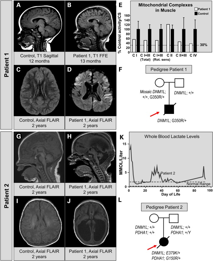
Clinical, neuroradiographic and molecular features of two patients with infantile encephalopathy and DNM1L variants. (A) MRI of a control patient at 12 months of age showing normal sagittal T1-weighted images. (B) MRI of Patient 1 at 13 months of age reveals hypoplasia of the corpus callosum and a simplified gyral pattern on T1 sagittal images. (C) MRI of control patient for suspected seizure showing a normal axial FLAIR sequence. (D) Axial FLAIR of Patient 1 at 2 years of age showing multi-focal areas of cortical signal abnormality with swelling and diffusion restriction. Diffuse cortical volume loss is also present. (E) Respiratory chain enzyme activities in muscle. The % control activity for Patient 1 (white bars) is shown for NADH:Ferricyanide dehydrogenase (CI), NADH:cytochrome C reductase total (CI+III), NADH:cytochrome C reductase Rotenone sensitive (CI+III), Succinate dehydrogenase (CII) and Cytochrome C oxidase (CIV). Values were normalized to citrate synthase activity. (F) Pedigree of the patient showing the WES data. Sanger verification was performed and low-level (6–8%) mosaicism was detected in the maternal sample. (G and I) A sagittal MRI in a newborn showing a normal T1 and FLAIR appearance. (H and J) MRI of Patient 2 at 4 days of age reveals ventriculomegaly, absence of the corpus callosum, volume loss and gyral simplification on T1 and FLAIR. (K) Whole blood lactate levels measured in a clinical lab over the time course of Patient 2's medical care at our institution. (L) Pedigree of Patient 2 showing the presence of two independent de novo events, one in DNM1L and one in PDHA1.

