Figure 6.
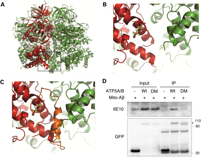
Binding of Aβ to ATP synthase. (A) Overall view of glycosylated ATP synthase complex. O-GlcNAcylation of ATP5A (red) on Thr432 is shown as a stick model. The neighboring β subunit (ATP5B) is colored in green. (B) Enlarged view of the O-GlcNAcylation site. (C) Molecular docking simulation between ATP synthase and Aβ. ATP5A, red; ATP5B, green; Aβ, orange. (D) Validation of Aβ binding on ATP synthase. Cells were transfected with control GFP vector or GFP-tagged WT or deletion mutant ATP5A/B (DM) in combination with mitochondria-targeted Aβ (mito-Aβ). After 48 h transfection, whole cell lysates were prepared and immunoprecipitated with anti-GFP antibody. The co-immunoprecipitated Aβ was detected with anti-Aβ mAb (6E10). Star indicates overexpressed protein.

