Figure 2.
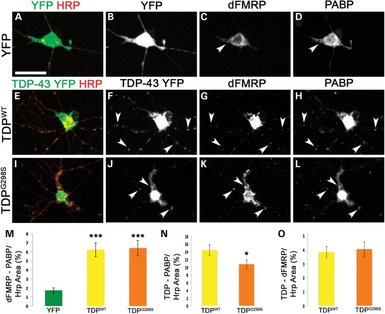
TDP-43, dFMRP and PABP colocalize in neuronal RNA granules in primary motor neurons. (A–D) Cytoplasmic YFP expressing cells immunostained with anti-GFP (A and B), anti-dFMRP (C), anti-PABP (D) and anti-Hrp (A) antibodies. (E–L) Primary neurons expressing TDP-43 YFP variants (TDPWT E-H, TDPG298S I–L) and immunostained with anti-GFP (E, F, I and J), anti-dFMRP (G and K), anti-PABP (H and L) and anti-HRP (E and I) antibodies. HRP labels neuronal membranes. Genotypes as indicated on the left, immunostains on the top. Arrowheads indicate select colocalizing TDP-dFMRP-PABP granules within neurites. (M) Quantification of co-localization between dFMRP and PABP normalized to HRP area. (N) Quantification of TDP-43 and PABP co-localization in normalized to HRP area. (O) Quantification of co-localization of TDP-43 and dFMRP normalized to HRP area. Student's T test was used to calculate P-values. *P < 0.05. Scale bars: (A) 12 μm.

