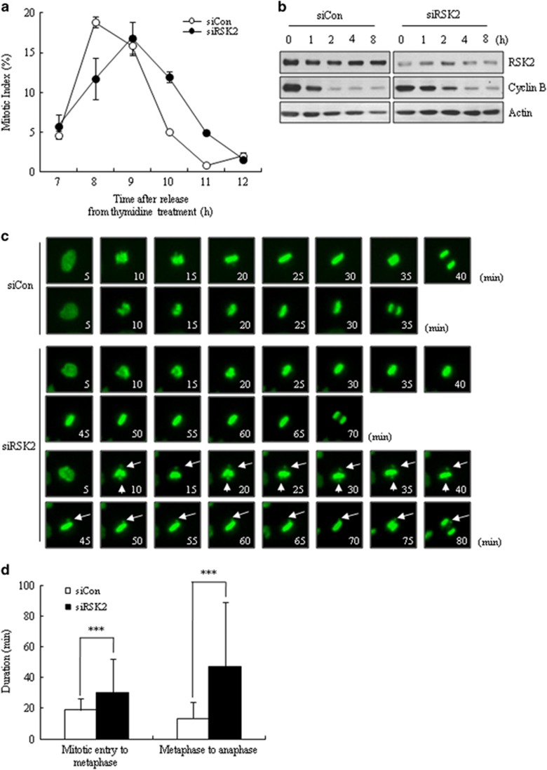
Figure 2.
Prolongation of mitotic duration by RSK2 depletion. (a) Mitotic index was scored at indicated time points after releasing from thymidine block (n=300). A representative data from three independent experiments are shown as mean±s.d. (b) Western blot analysis of cyclin B degradation was performed after releasing from nocodazole treatment (100 ng ml−1) for 16 h. (c) Time-lapse microscopy imaging of HeLa cells stably expressing a chromatin marker (H2B–green fluorescent protein; green); rounding up is marked as 0 min. Arrow: unaligned chromosomes. ***P<0.001 by Student's t-test. (d) Mitotic duration was measured from time-lapse images (n=100). Mitotic entry time was set as the time of rounding up of the cell. Results are shown as the mean±s.d. from three independent experiments. ***P<0.001 by Student's t-test.

