Figure 3.
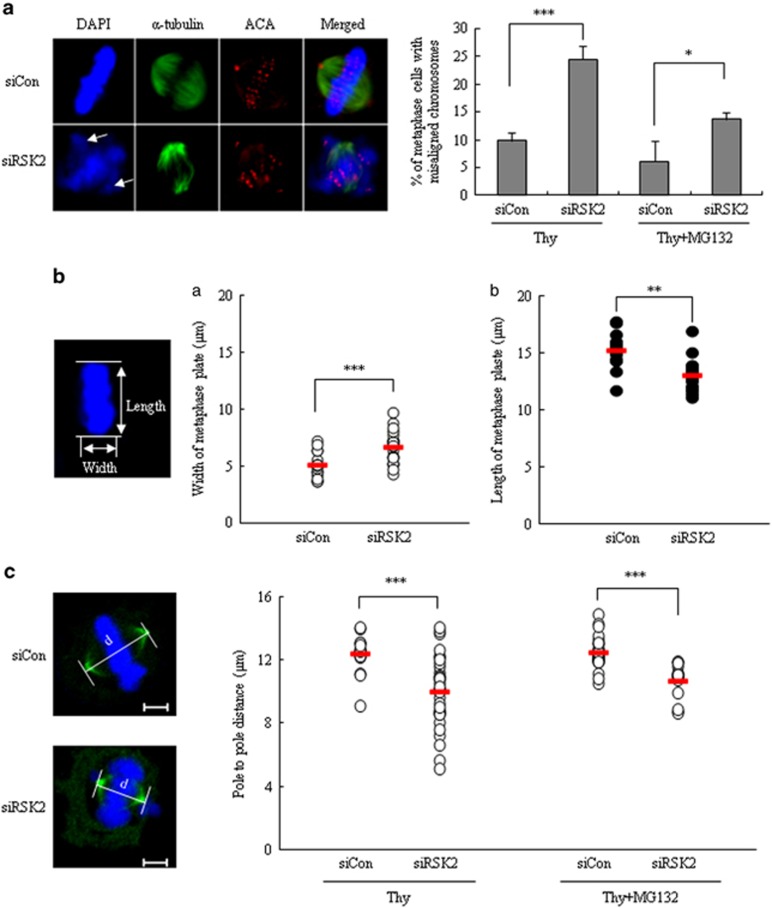
Defects of the chromosome congression and alignment in RSK2-depleted cells. (a) Unaligned chromosomes of control and RSK2-depleted cells were measured 8 h after releasing from thymidine block with or without 2 h more incubation with MG132. MG132 was used to give enough time for chromosome congression. Images were representative control (upper left) or RSK2-depleted HeLa cells (lower left) stained for ACA (red), α-tubulin (green) and DNA (blue). Arrow: unaligned chromosomes. Percentage of metaphase cells with unaligned chromosomes. Results are shown as the mean±s.d. from three independent experiments. *P<0.05, ***P<0.001 by Student's t-test (n=100). (b) The width (a) and length (b) of congressed body of chromosomes on metaphase plate were measured 8 h after releasing from thymidine block. A representative data from three independent experiments are shown as mean±s.d. **P<0.005, ***P<0.001 by Student's t-test (n=20). Representative image of metaphase plate was stained with 4′,6-diamidino-2-phenylindole. (c) Pole-to-pole distance of metaphase cells (left panel) was measured at 8 h after releasing from thymidine block with or without 2 h more incubation with MG132. A representative data from three independent experiments are shown as mean±s.d. ***P<0.001 by Student's t-test (n=16–36). Representative images of control (upper) or RSK2-depleted HeLa cells (lower) stained for γ-tubulin (green) and DNA (blue). Scale bar, 5 μm.

