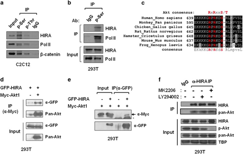
Figure 1.
HIRA interacts with Akt1. (a, b) Whole-cell extracts prepared from C2C12 (a) or HEK293T (b) cells were subjected to IP using anti-p-Ser, anti-p-Thr, and control IgG antibodies, they were then probed with anti-HIRA (WC119) to detect phospho-HIRA. Two phospho-proteins, RNA polymerase II and β-catenin, were used as positive controls. (c) Sequences potentially targeted by Akt (RxRxxS/T) in vertebrate homologues were aligned. (d, e) GFP-HIRA was co-expressed with Myc-Akt1 in 293T cells. Whole-cell extracts were subjected to IP and immunoblotted using the indicated antibodies. Five percent of the input sample was loaded in parallel. *Non-specific background. (f) 293T cells were treated with MK2206 (10 μM) or LY294002 (10 μM) and the whole-cell extracts were subjected to IP using anti-HIRA(WC15) or IgG. Anti-pan-Akt, p-Akt(S473), or HIRA(WC119) antibodies were used for immunoblotting. TBP (TATA-binding protein) was used as the loading control. GFP, green fluorescent protein.

