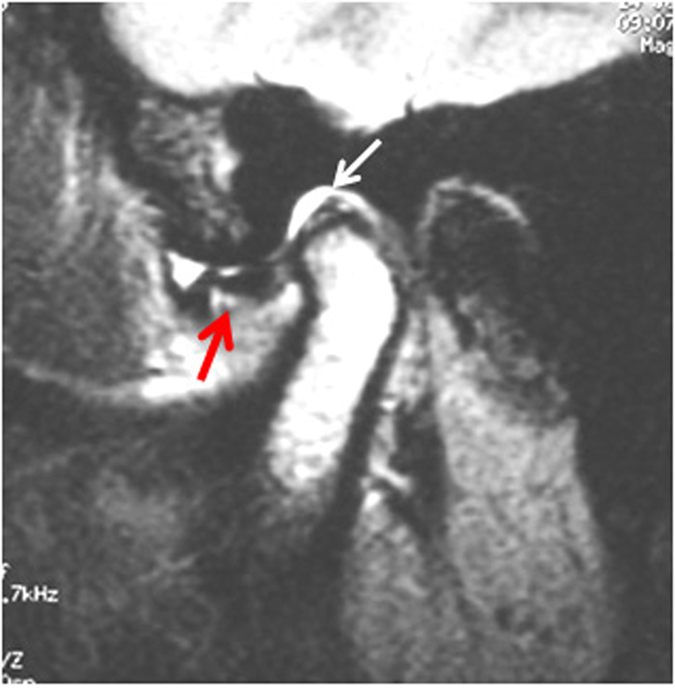
Figure 1. MRI showed acute phase of ATDD, disc displaced in front of the condyle (red arrow) with normal shape and length.

The posterior band was disrupted and elongated with or without effusion (white arrow).

The posterior band was disrupted and elongated with or without effusion (white arrow).