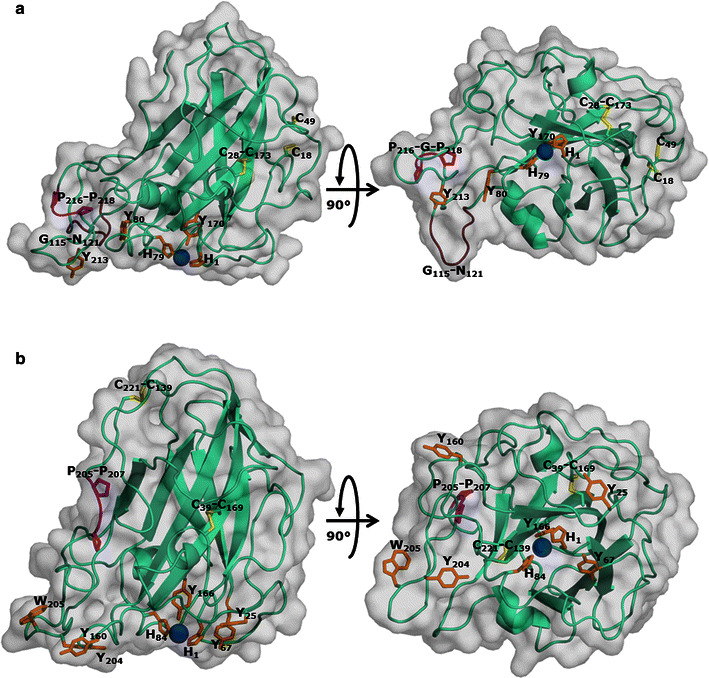
Fig. 3.

Structural models of MtLPMO9B and MtLPMO9C. The structural model of a MtLPMO9B was generated based on the available structure of NcLPMO9C from Neurospora crassa [25] (PDB entry: 4D7U). The amino acid sequence identities of MtLPMO9B and MtLPMO9C compared to NcLPMO9C were 41 and 46 %, respectively. The copper ion (blue) is coordinated by His1, His 79 and Tyr170 (orange). A disulfide bridge is located between Cys28 and Cys178 (yellow) and it is likely that the neighboring Cys18 and Cys49 form a second disulfide bridge. The structural model of b MtLPMO9C was, like MtLPMO9B, generated based on NcLPMO9C [25] (PDB entry: 4D7U). The copper ion (blue) is coordinated by His1, His84 and Tyr166 (orange). MtLPMO9C contains two disulfide bridges between Cys39 and Cys169, and Cys139 and Cys221 (yellow). The highly conserved Gly-Pro-Gly triad (magenta) of MtLPMO9B and MtLPMO9C is located between Pro216 and Pro218, and Pro205 and Pro207, respectively. MtLPMO9B contains, unlike MtLPMO9C, an additional loop between Gly-115 and Asn121 (brown)
