Fig. 4.

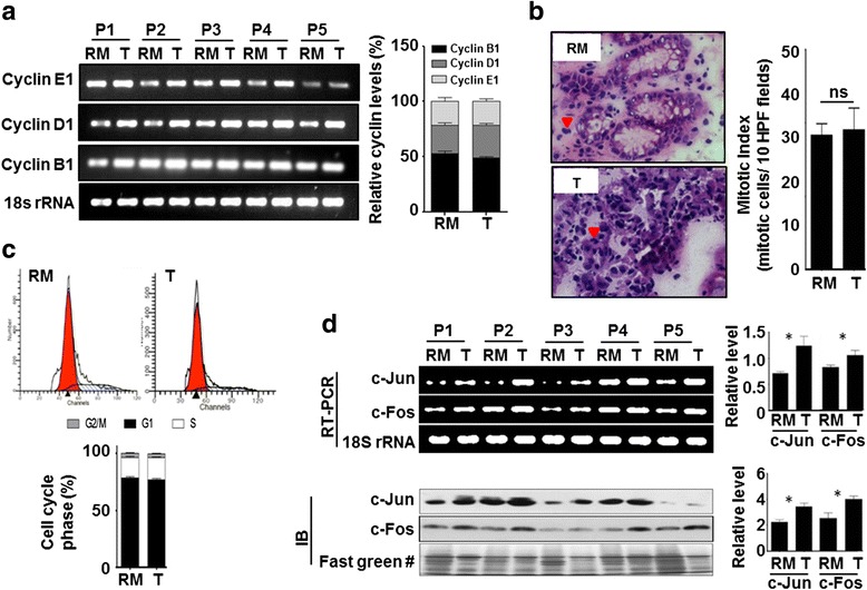
Association of cell cycle profile with GC tumor and resection margin tissues. a RT-PCR analysis shows a high mRNA level of cyclin B1, D1, and E1 in tumor than resection margin tissues (left panel). mRNA level of cyclins were normalized with 18S rRNA, combined % was calculated for each cyclin in tumor and resection margin tissues, and their relative % levels were compared showing no difference in cell cycle profile of tumor and resection margin tissues (right panel). b Arrow heads showing mitotic cells in H&E-stained resection margin and tumor tissue section image (×40, left panel). On H&E-stained tissue sections, mitotic index was calculated for paired samples (n = 40), comparison between tumor and resection margin tissues showing no significant difference (right panel). c Flow cytometry-based cell cycle profile showed most of the cells of both tumor and resection margin tissues are in G1 phase (upper panel). Comparison of mean % (n = 10) of G2/M, G1, and S phases of cell cycle showed no difference in cell cycle profile of tumor and resection margin tissues (lower panel). d RT-PCR (left upper panel) and immunoblot (left lower panel) analyses showed a high level of immediate early genes, c-jun and c-fos, in tumor than resection margin tissues. After normalization, comparison of relative level also showed significant increase of c-jun and c-fos in tumor tissues, both at transcript (right upper panel) and protein (right lower panel) level. GC gastric cancer, RM resection margin either PRM or DRM with maximum distance from the site of the tumor, T tumor, P patient. #Fast green-stained PVDF membrane used in Fig. 5a as well. Statistical tests are done by using Wilcoxon matched pairs test. *p < 0.05, **p < 0.01, ***p<0.001
