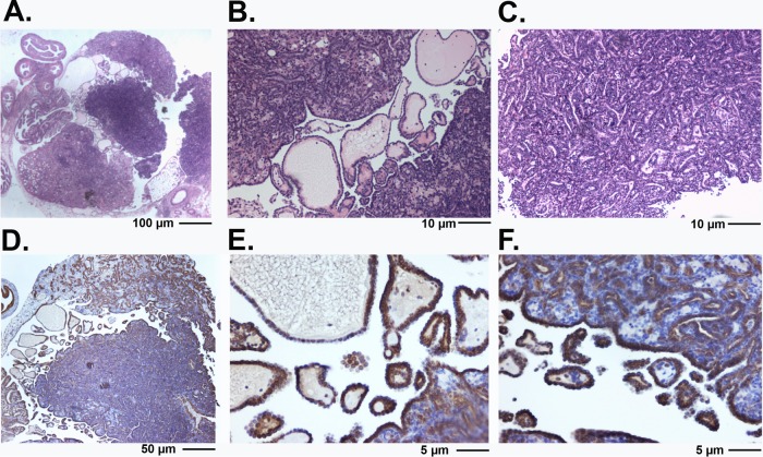
FIG 3.
Morphology of ovarian tubular adenomas in Wv/Wv; p27+/− mice. (A) H&E staining of a large ovarian tubular adenoma from an 8-month-old Wv/Wv; p27+/− mouse. The tumor is papillary and resembles a serous borderline ovarian tumor. (B) Higher magnification of the tumor in panel A showing the presence of papillary adenoma morphology. (C) Higher magnification of the tumor in panel A showing the crowding of epithelial tubular gland structures. (D) The same tumor stained with cytokeratin 8. (E and F) Cytokeratin 8 staining of the tumor shown at higher magnification.

