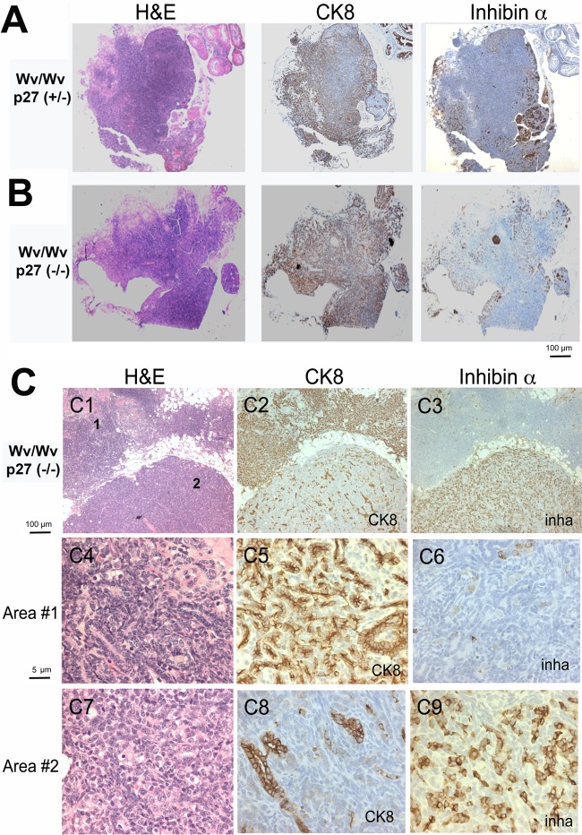
FIG 4.
Inverse correlation between the presence of granulosa cells and epithelial lesions in Wv/Wv; p27+/− and Wv/Wv; p27−/− ovaries. (A) Representative tissue sections stained with H&E, the granulosa cell marker inhibin α, and the epithelial marker CK8 in adjacent sections of ovarian tubular adenomas from a 12-month-old Wv/Wv; p27+/− mouse. (B) Staining of a representative ovarian tubular adenoma from a 12-month-old Wv/Wv; p27−/− mouse. (C) An ovarian tubular adenoma from an 8-month-old Wv/Wv; p27−/− mouse was analyzed in more detail (C1 to C3) for H&E, CK8, and inhibin α (inha) staining. Images at higher magnification (numbered in panel C1) are shown for area 1, which is CK8 positive (C4, H&E; C5, CK8; C6, inhibin α), and area 2, which is inhibin α positive (C7, H&E; C8, CK8; C9, inhibin α).

