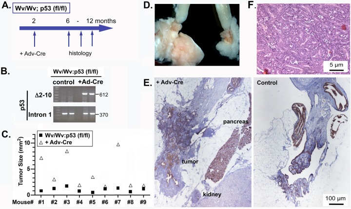
FIG 5.
Ovarian tumors in Wv/Wv; p53fl/fl; Adv-Cre mice. (A) Schematic of the experimental protocol, in which 2-month-old Wv/Wv; p53fl/fl female mice were injected with Adv-Cre in the ovarian bursa of one side; the mice were then analyzed 4 to 10 months later (at 6 to 12 months of age). (B) DNA extracted from the ovarian or tumor tissues was used for PCR genotyping for the wild-type p53 allele and the delta floxed (df) allele (with deletion of exons 2 to 10). Adv-LacZ or Adv-GFP was used in the controls. (C) Tumor sizes were quantified based on a histological slide near the widest cross section of the ovary/tumor. The ovary/tumor pairs from injected and uninjected control sides were plotted for comparison. The difference between the two groups was statistically significant based on paired two-tailed Student t tests (P < 0.001). (D) Typical gross morphology of ovaries and uterine horns of a 10-month-old Wv/Wv; p53fl/fl; Adv-Cre mouse. The left ovary was injected with Adv-Cre. (E) Sections of a typical pair from uninjected or Adv-Cre-injected ovarian tissue or derived tumors were stained with CK8. The control (uninjected) ovary is shown for comparison. (F) H&E staining showing the tumor cell morphology of the ovarian adenoma from a 10-month-old Wv/Wv; p53fl/fl; Adv-Cre mouse.

