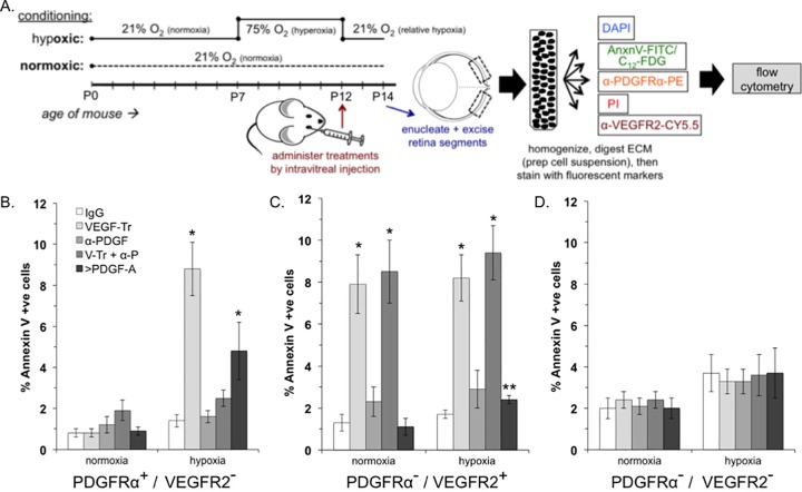
FIG 3.
VEGF promoted the viability of PDGFRα+/VEGFR2− retinal cells in mice enduring relative hypoxia. (A) Overview of the experimental procedure, which is described in detail in Materials and Methods. (B to D) Percentages of FITC-annexin V-positive (apoptotic) cells that were PDGFRα+/VEGFR2− (B), PDGFRα−/VEGFR2+ (C), or PDGFRα−/VEGFR2− (D). The data are reported as the means ± the SD from five eyes (n = 5), comprising 50,000 events in total. Significant differences compared to IgG for each condition was determined using a paired t test, where a single asterisk denotes P < 0.01 and double asterisks denote P < 0.05.

