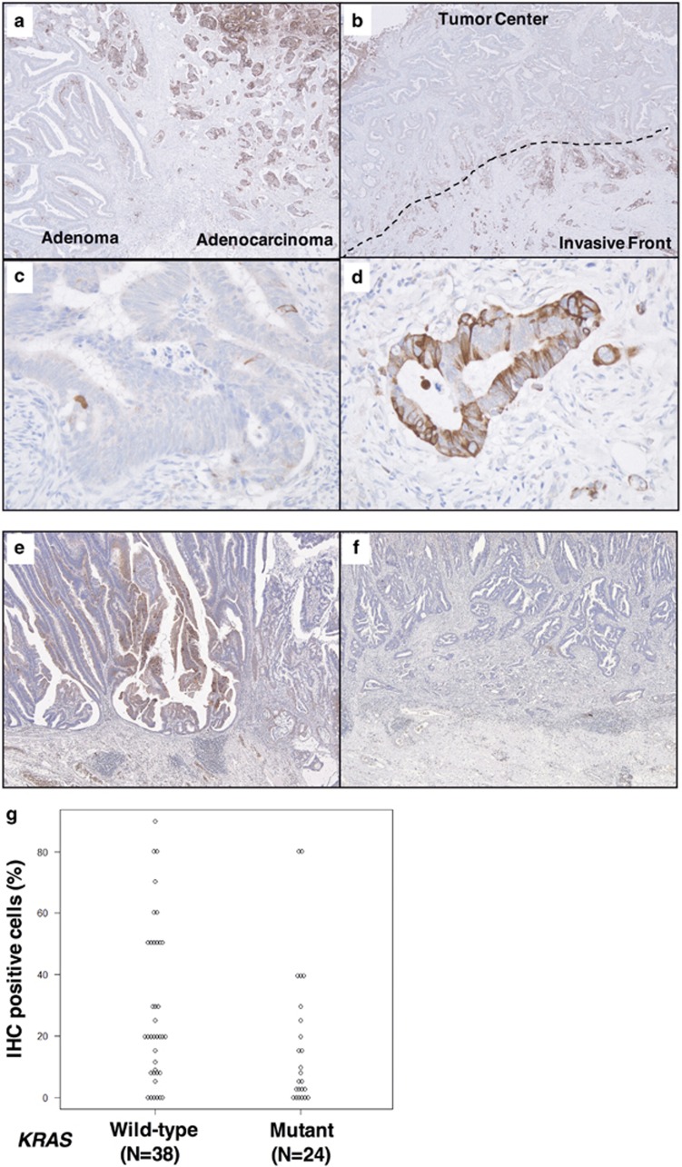
Figure 2.
Immunohistochemical analyses of RCAN2 in surgical specimens from patients with colorectal cancer (CRC). Representative images of immunohistochemical staining (IHC) at the border between adenoma (right, in a) and adenocarcinoma (left, in a), and the intratumoral distribution of RCAN2 expression (b). Magnified views at the tumor center and the invasive front of tumors (c, d, respectively). Representative IHC images of KRAS wild-type and KRAS-mutated human CRC tissue samples (e, f, respectively). Comparison of IHC-positive cells in the carcinomatous area of KRAS wild-type and KRAS-mutated CRC indicated lower expression of RCAN2 in KRAS-mutated CRC (P=0.0447, g).

