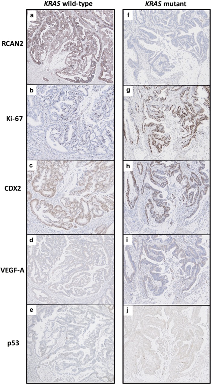
Figure 3.
Immunohistochemical analyses of RCAN2 and other markers at invasive front of cancer. Representative IHC images of KRAS wild-type (a–e) and KRAS-mutated (f–j) human CRC tissue samples, stained with anti-RCAN2 (a, f), anti-Ki-67 (b, g), anti-CDX2 (c, h), anti-VEGF-A (d, i), and anti-p53 (e, j) antibodies.

