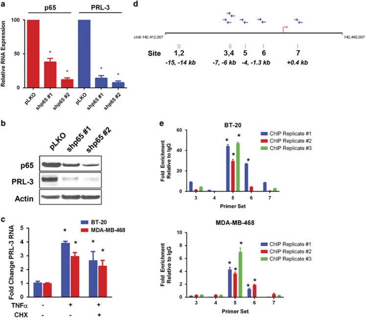
Figure 3.
The NF-ĸB transcription factor subunit, p65, binds the PRL-3 promoter and regulates PRL-3 expression. (a) BT-20 RNA levels as assessed by qRT–PCR for p65 (red) and PRL-3 (blue) following p65 knockdown using two different shRNA clones (shp65 #1 and shp65 #2). (b) Immunoblot analysis of p65 and PRL-3 protein levels following p65 knockdown in BT-20 cells. (c) Fold change in PRL-3 RNA in BT-20 (blue) and MDA-MB-468 (red) cells as determined by qRT–PCR after 6 h treatment with TNFα (20 ng/ml) or TNFα (20 ng/ml) and cycloheximide (CHX, 100 nM). (d) Schematic of the human PRL-3 promoter locus. Seven potential NF-ĸB-binding sites indicated with green hash marks and numbered, with the position of each site with respect to the transcription start site (red arrow) indicated. Blue arrows represent qRT–PCR oligonucleotide primers designed to recognize the respective binding sites. (e) Results of three independent replicate chromatin immunoprecipitation experiments performed in BT-20 and MDA-MB-468 cells following 30 min treatment with 20 ng/ml TNFα. Data are represented as fold enrichment relative to IgG control. Data represented are the mean±s.d. of three independent experiments. *P<0.05 as determined by Student t-test.

