Figure 1.
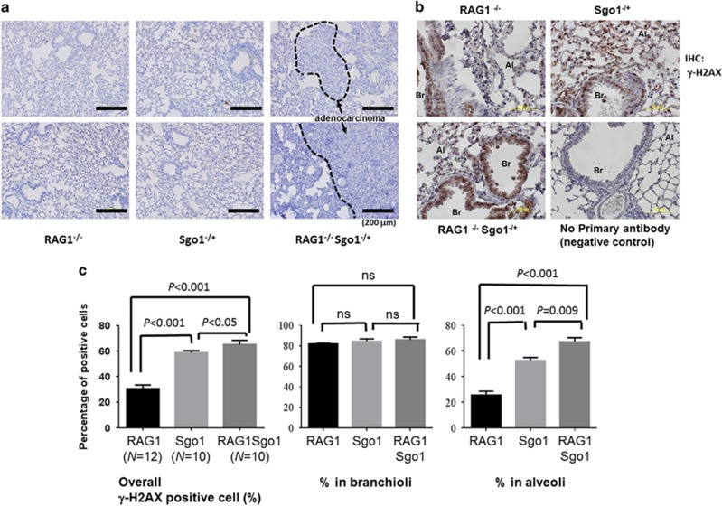
RAG1−/− Sgo1−/+ mice developed lung adenocarcinomas. (a) RAG1−/− Sgo1−/+ mice developed lung adenocarcinomas with the appearance of bronchioloalveolar in-type (that is, cuboidal cells lining the alveolar septa and projecting into alveolar spaces). RAG1−/− and most Sgo1−/+ did not show significant histopathological changes, with some exceptions in Sgo1−/+ that developed adenocarcinomas in smaller sizes. (b) To assess DNA double-strand break, IHC for γ-H2AX was performed. Percentages for γ-H2AX positive cells were high in Branchioli, but varied between Sgo1−/+ and Sgo1+/+ backgrounds in alveoli. Al, alveole; Br, branchiole. (c) Rag1−/−Sgo1−/+ showed highest percentage of γ-H2AX positive cells. However, signals at branchioli showed little difference (NS, nonsignificant). The difference between Sgo1−/+ and Sgo1+/+ backgrounds were evident only in alveoli.

