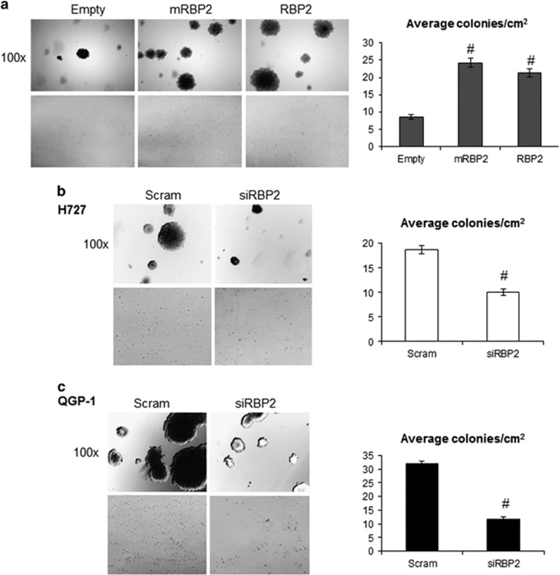
Figure 6.
Colony formation with RBP2 overexpression and knockdown. (a) βlox5 cells stably expressing either empty vector (Empty), enzymatically dead mutant RBP2 (mRBP2), or wild-type RBP2 (RBP2) were used for colony formation assays and (b) H727 and (c) QGP-1 cells were transiently transfected with either a scrambled siRNA (Scram) or an RBP2 siRNA (siRBP2) and followed 24 hrs later by colony formation assays. Colonies were stained with crystal violet after 3 weeks and pictures were taken at × 100 magnification or with a standard hand held camera. In addition, colonies present in 25 × 1 cm2 fields were counted and an average number of colonies per cm2 was calculated for each condition. (#P<0.005).

