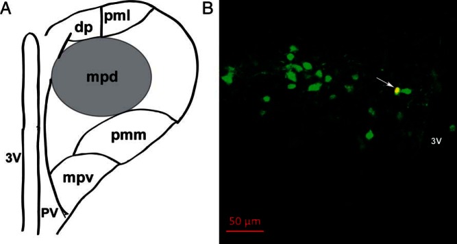
Figure 1.
Identification of recorded CRH neurons in hypothalamic paraventricular nucleus (PVN) region. A, Schematic representation of the mouse PVN, highlighting the anatomical location of putative neuroendocrine CRH neurons in the mpd region (black circle), located between the third ventricle and lateral cluster of large neurons. B, Confirmation of the neurochemical profile of recorded CRH neurons after patch clamp recording by intracellular dye labeling (Alexa Fluor 594, added 0.1% intracellularly into internal solution) followed by immunohistochemical analysis. A representative image is shown as an example (yellow; see arrow) of an identified CRH-immunopositive (Alexa Fluor 633, green) neuron in the PVN region of a coronal mouse brain slice, with its recorded location labeled with Alexa Fluor 594 (red). Scale bar, 50 μm. Abbreviations: dp, dorsal parvocellular division; mpd, dorsal portion of the medial parvocellular division; mpv, ventral portion of the medial parvocellular division; pmm, medial portion of posterior magnocellular division; pml, lateral portion of posterior magnocellular division; pv, periventricular parvocellular division; 3V, third ventricle.

