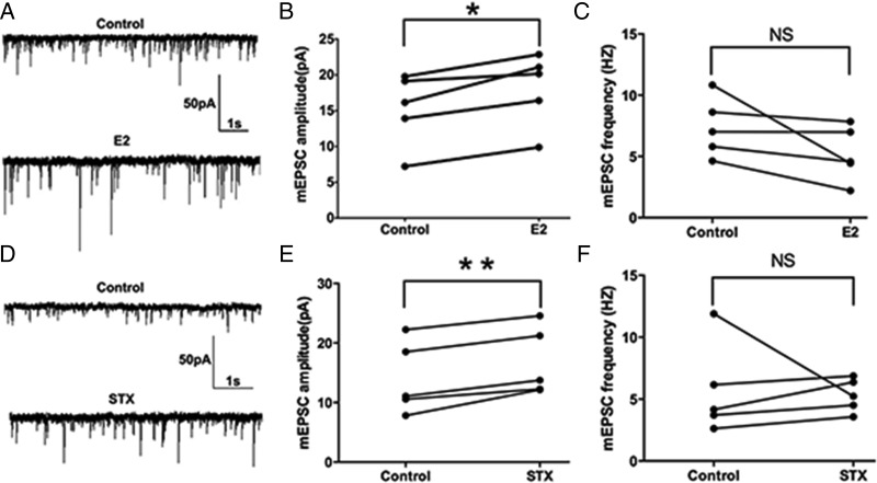
Figure 5.
E2 and STX rapidly potentiated the amplitude of miniature EPSCs (mEPSC) in PVH CRH neurons. A, An example of mEPSC during control conditions and perfusion of E2 (10 min, 100nM). B, Average mEPSC amplitude increased after perfusion of E2. C, Average mEPSC frequency did not change after E2 perfusion. D, An example of mEPSC during control conditions and perfusion of STX (10 min, 100nM). E, Average mEPSC amplitude increased after perfusion of STX. F, Average mEPSC frequency did not change after STX perfusion. Scale bar shows a 50-pA amplitude and 1-second duration. Data were analyzed by a paired Student's t test (*, P < .05; **, P < .01). NS, no significant difference (P > .05).

